Anatomy Muscles Pelvis - The Pelvic Floor Structure Function Muscles Teachmeanatomy - Pelvic outlet/ inferior aperture of pelvis separates pelvic cavity from perineum.its boundaries are as follows:.
Anatomy Muscles Pelvis - The Pelvic Floor Structure Function Muscles Teachmeanatomy - Pelvic outlet/ inferior aperture of pelvis separates pelvic cavity from perineum.its boundaries are as follows:.. The anterior muscles of the femur extend the lower leg but also aid in flexing the thigh. Figure 11.29 hip and thigh muscles the large and powerful muscles of the hip that move the femur generally originate on the pelvic girdle and insert into the femur. The pelvis's frame is made up of the bones of the pelvis, which connect the axial skeleton to the femurs, and therefore acts in weight bearing of the upper body. Tightness of these ligaments and tendons can cause hip instability and pain. See more ideas about anatomy, pelvic floor, pelvic floor dysfunction.
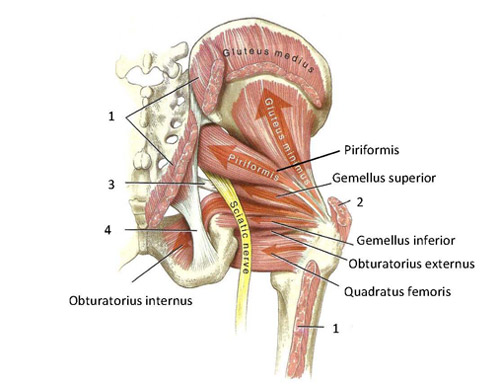
See more ideas about anatomy, pelvic floor, pelvic floor dysfunction. Rather, their function is primarily to stabilize the sacroiliac and symphysis pubis joints as well as to create a stable floor for the visceral contents of the abdominopelvic cavity. Those two muscles make up part of the walls of the pelvis. The large intestine ends in the rear of the pelvis at the anus, a sphincter muscle that controls the disposal of solid waste. Attached to the pelvis are muscles of the buttocks, the lower back, and the thighs.

Included in this group are the adductor longus, adductor brevis, adductor magnus, pectineus, and gracilis muscles.
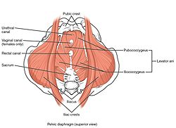
Enumerate the muscles of true pelvis. The hip has several ligaments connecting the femur to the pelvis and tendons connecting the bones to many surrounding muscles. The floor of the pelvis is made up of the muscles of the pelvis, which support its contents and maintain urinary and faecal continence. Flashcard anatomy on the muscles of the pelvis. The muscles of the pelvic floor are collectively referred to as the levator ani and coccygeus muscles. The pelvis also houses the reproductive organs, which have their own muscles. The gluteus medius and minimus are also important stabilizers of the hip joint and help to keep the pelvis level as we walk. The pelvic girdle, also known as the hip bone, is composed of three fused bones: The it band is a common cause of lateral (outside) hip, thigh, and knee pain. Gracilis is the most superficial and medial muscle of the group. These muscles move the thigh toward the body's midline. The pelvic floor separates the pelvic cavity above from the perineum below. It can be divided into the greater pelvis and the lesser pelvis.
At its insertion, the tendon of the sartorius muscle lies anterior and the tendon of semitendinosus lies posterior. The muscles of true pelvis are: The pelvic floor muscles provide foundational support for the intestines and bladder. The pelvic girdle, also known as the hip bone, is composed of three fused bones: The thigh bone or femur and the pelvis join to form the hip joint.

These muscles, including the gluteus maximus and the hamstrings, extend the thigh at the hip in support of the body's weight and propulsion.
See more ideas about anatomy, pelvic floor, pelvic floor dysfunction. The levator ani muscles are the largest group of muscles in the pelvis. The pelvic floor muscles provide foundational support for the intestines and bladder. Pelvic outlet/ inferior aperture of pelvis separates pelvic cavity from perineum.its boundaries are as follows:. The hip has several ligaments connecting the femur to the pelvis and tendons connecting the bones to many surrounding muscles. Figure 11.29 hip and thigh muscles the large and powerful muscles of the hip that move the femur generally originate on the pelvic girdle and insert into the femur. Flashcard anatomy on the muscles of the pelvis. It can be divided into the greater pelvis and the lesser pelvis. This mri male pelvis axial cross sectional anatomy tool is absolutely free to use. Muscles of the pelvic floor do not cross from the pelvis to another body part; The floor of the pelvis is made up of the muscles of the pelvis, which support its contents and maintain urinary and faecal continence. The ilium, ischium and the pubic bone. The adductor muscle group, also known as the groin muscles, is a group located on the medial side of the thigh.
Lying exposed between the protective bones of the superiorly located ribs and the inferiorly located pelvic girdle, the muscles of this region play a critical role in protecting the delicate vital organs within the abdominal cavity. It can be divided into the greater pelvis and the lesser pelvis. Tightness of these ligaments and tendons can cause hip instability and pain. Gracilis is the most superficial and medial muscle of the group. Included in this group are the adductor longus, adductor brevis, adductor magnus, pectineus, and gracilis muscles.
:background_color(FFFFFF):format(jpeg)/images/library/11895/male-pelvic-viscera-and-perineum_english.jpg)
The muscles of the pelvic floor are collectively referred to as the levator ani and coccygeus muscles.
Included in this group are the adductor longus, adductor brevis, adductor magnus, pectineus, and gracilis muscles. The ilium, ischium and the pubic bone. Support of abdominopelvic viscera (bladder, intestines, uterus etc.) through their tonic contraction. The gluteus medius and minimus are also important stabilizers of the hip joint and help to keep the pelvis level as we walk. It is usually divided into two separate anatomic regions: The hip joint is a ball and socket synovial joint, formed by an articulation between the pelvic acetabulum and the head of the femur. The hip has several ligaments connecting the femur to the pelvis and tendons connecting the bones to many surrounding muscles. Gracilis is the most superficial and medial muscle of the group. The anterior muscles of the femur extend the lower leg but also aid in flexing the thigh. The pelvic floor muscles provide foundational support for the intestines and bladder. They form a large sheet of skeletal muscle that is thicker in some areas than in others. Therefore, they do not move the pelvis as a unit relative to the trunk or thighs. See more ideas about anatomy, pelvic floor, pelvic floor dysfunction.
Komentar
Posting Komentar